Lung pet scan: purpose, procedure, and preparation.
Positron Emission Tomography Wikipedia

Lung Pet Scan Purpose Procedure And Preparation
A positron emission tomography (pet) scan is an imaging test that helps reveal how your tissues and organs are functioning. a pet scan uses a radioactive drug (tracer) to show this activity. this scan can sometimes detect disease before it shows up on other imaging tests. neck mri pelvic mri pacemaker cardiac pacemaker implantation pet scan pet scan (chest to head neck) pet scan (skull to mid-thigh) pet scan brain pet scan heart whole body pet scan reflux surgery gastric cardioplasty septoplasty surgery septoplasty sleep lithotripsy (kidney stone removal) mammogram mra mri pacemaker pet scan reflux surgery septoplasty surgery sleep study spinal cord A pet scan (also known as positron emission tomography and pet/ct) is a type of imaging study that can show doctors what’s happening in your body and how it’s working. it’s different from an x-ray,. Positron emission tomography, also called pet imaging or a pet scan, is a diagnostic examination that involves getting images of the body based on the detection of radiation from the emission of positrons. positrons are tiny particles emitted from a radioactive substance administered to the patient. patient safety tips prior to the exam.
Pet Scan Cancer Staging And Treatment Verywell Health
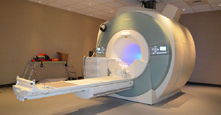
A pet (positron emission tomography) scan is a type of imaging test that uses radioactive glucose (radiotracer) to see where cancer cells may be. since cancer cells intake more glucose than normal cells, injecting some glucose into a vein can reveal where cancerous cells are in the pet scan body when scanned and examined with computerized images. Pet scans are performed on an outpatient basis, most commonly in the nuclear medicine imaging unit of a hospital or in a dedicated facility. the room itself is called either the scanning room or procedure room. the pet scanner is a large machine with a doughnut-shaped hole in the center, similar to a ct or mri unit. Definition a positron emission tomography (pet) scan is an imaging test that allows your doctor to check for diseases in your body. the scan uses a special dye containing radioactive tracers. these.  compare pet scan cost welcome to comparepetscancost. com where you can: learn about pet (positron emission tomography) scan procedures determine average.
Radiology Mri Scans Petct Scans Information
A pet scan detects changes in cellular function how your cells are utilizing nutrients like glucose and oxygen. a ct scan uses a combination of x-rays and computers to give the radiologist a non-invasive way to see inside the body. when these two scans are fused together, the san diego imaging radiologists can view metabolic changes in the. Positron emission tomography (pet) is a sophisticated medical imaging technique. it uses a radioactive tracer to pinpoint differences in tissues on the molecular level. a whole-body pet scan can.

Pet Scan Definition Purpose Procedure And Results
contact us diagnostic radiology ultrasound directory radiology: mri scans & pet/ct scans information looking for collecting the information on the know about various procedures and therapies, such as pet scans ct scans pet scan and even mri scans that help provided in this website tells you how various pet scans ct scans or mri scans and provides some Provides whole-body positron emission tomography (pet) scan services to physicians and patients in the bay area for oncology, cardiology and neurology. (san jose, california). A new study led by researchers at the ucla jonsson comprehensive cancer center helps identify which patients with prostate cancer will benefit most from the use of prostate-specific membrane antigen pet imaging,.
Pet scan is an imaging technique that uses a radioactive tracer to locate tissue differences at a molecular level. a lung pet scan is used to take images of the lungs and detect whether lung. Positron emission tomography (pet) is a type of imaging technology used to evaluate how your tissues and organs work at the cellular level. it involves the injection of a short-acting radioactive substance, known as a radiotracer, which is absorbed by biologically active cells. A positron emission tomography (pet) scan produces images of your organs and tissues at work. the test uses a safe injectable radioactive chemical called a radiotracer and a device called a pet scanner. the scanner detects diseased cells that absorb large amounts of the radiotracer, which indicates a potential health problem. A pet scan (also known as positron emission tomography and pet/ct) is a type of imaging study that can show doctors what’s happening in your body and how it’s working. it’s different from an.
A positron emission tomography, also known as a pet scan, produces 3-d color images of the processes within the human body. pet scans are often used to diagnose a condition or track how it is. Positron emission tomography (pet) scans are a type of imaging test your doctor uses to identify possible diseases at the cellular level. the scan allows physicians to evaluate oxygen intake, blood flow and metabolism to create a clearer picture of systemic diseases like cancer, brain disorders and heart problems. Further increasing the availability of pet imaging is a technology called gamma camera systems (devices used to scan patients who have been injected with small amounts of radionuclides and currently in use with other nuclear medicine procedures). these systems have been adapted for use in pet scan procedures.
Provides a layman''s pet scan introduction, explaining the indications, uses and how it works, and what to expect during the scan. also introduces pet scans. Positron emission tomography (pet) is a functional imaging technique that uses radioactive substances known as radiotracers to visualize and measure changes in metabolic processes, and in other physiological activities including blood flow, regional chemical composition, and absorption. different tracers are used for various imaging purposes, depending on the target process within the body. Positron emission tomography (pet) is a functional imaging technique that uses radioactive substances known as radiotracers to visualize and measure changes in metabolic processes, and in other physiological activities including blood flow, regional chemical composition, and absorption. different tracers are used for various imaging purposes, depending on the target process within the body.
A positron emission tomography, also known as a pet scan, uses radiation to show activity within the body on a cellular level. it is most commonly used in cancer treatment, neurology, and. A pet scan is a painless procedure that helps detect disease sooner than ct or mri images. a combination pet/ct scan provides a more detailed look at organs and tissue. pet scans can ensure an accurate diagnosis and help your healthcare provider develop an effective treatment plan.
What is a pet scan? a pet scan uses a radiotracer to measure things like blood flow, oxygen use and sugar metabolism. a pet scan shows how your tissues and organs are functioning. it also can let you and your doctors know if cancer treatment is working. follow your pet scan prep for best results. Positron emission tomography, also called petimaging or a pet scan, is a diagnostic examination that involves getting images of the body based on the detection of radiation from the emission of positrons. positrons are tiny particles emitted from a radioactive substance administered to the patient. patient safety tips prior to the exam.
wrist, hand (upper extremity) neck mri pelvic mri pet scan pet scan (chest to head neck) pet scan (skull to mid-thigh) pet scan brain pet scan heart whole body pet scan A positron emission tomography (pet) scan is an imaging test that uses a special dye with radioactive tracers. the tracers are either swallowed, inhaled, or injected into your arm. they help your.

